Current participating members in the ENIGMA-Stroke Recovery working group: Member List

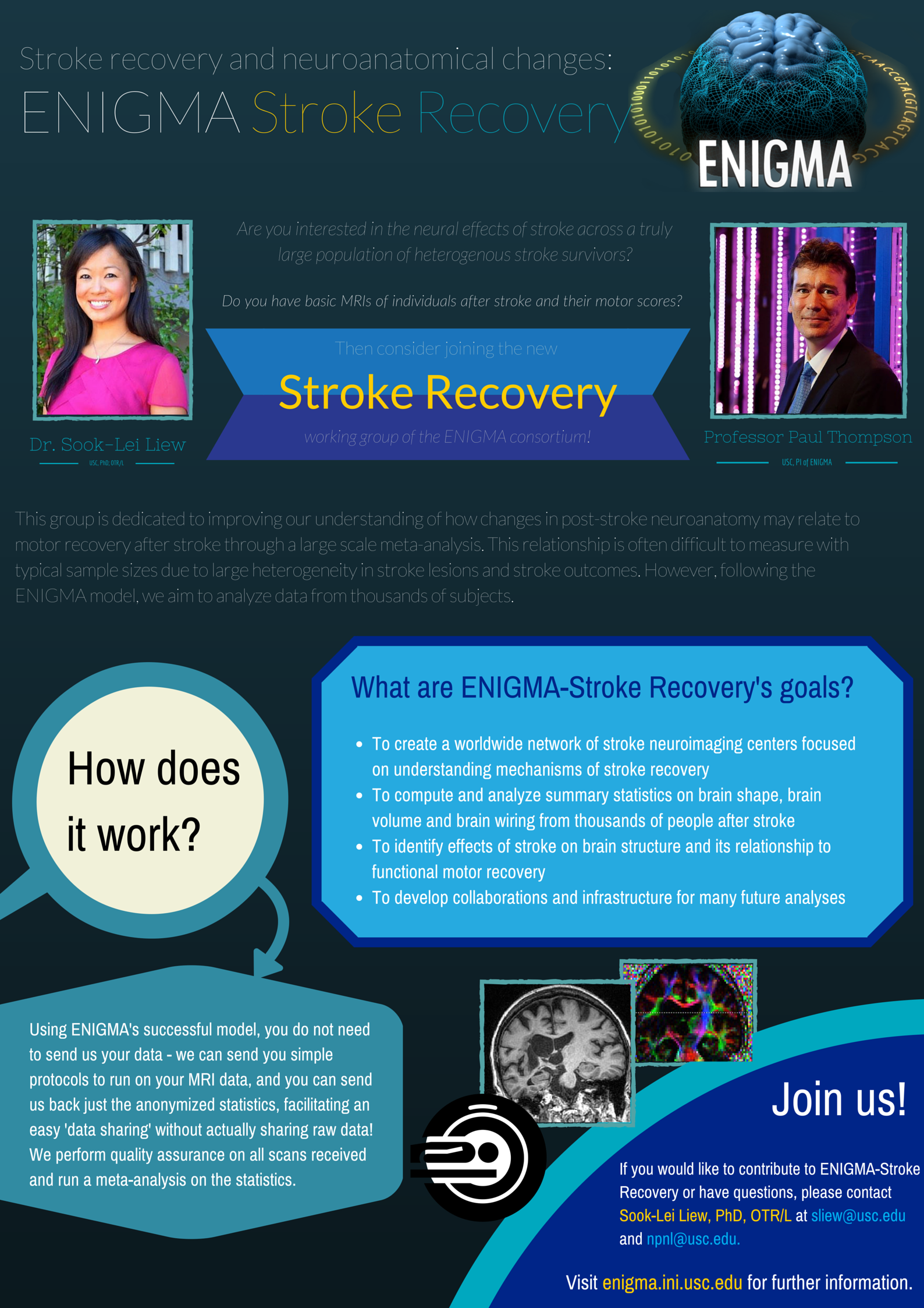
ENIGMA-Stroke Recovery is a working group dedicated to improving our understanding of how changes in the brain after stroke relate to functional outcomes and recovery. This group is actively conducting meta- and mega-analyses of existing MRI data in adults who have had a stroke. The main goals of ENIGMA-Stroke include:
-
To create a worldwide network of stroke neuroimaging centers focused on understanding mechanisms of stroke recovery
-
To compute and analyze metrics of brain shape, brain volume, brain wiring and brain function from thousands of people after stroke
-
To identify structural and functional differences that occur after stroke in brain shape, brain volume, brain wiring and brain function and explore the relation between these measures and functional outcomes and/or recovery
-
To develop collaborations and infrastructure for novel stroke brain-behavior analyses
Although initial work from this worldwide partnership focused on examining sensorimotor outcomes after stroke, our growing membership is also interested in examining neural associations with post-stroke on cognition, mood, language, and more, as well as the effects of therapeutic approaches, exercise and sleep on stroke outcomes. We are also interested in multi-modal imaging, genetics, and additional neurobiological metrics of health.
We are happy to welcome new cohorts at any time! If you would like to contribute to ENIGMA-Stroke, please contact Sook-Lei Liew.
ENIGMA Stroke Recovery Papers:
- ENIGMA Stroke Recovery Overview:
Liew, S. L., Zavaliangos‐Petropulu, A., Jahanshad, N., Lang, C. E., Hayward, K. S., Lohse, K. R., ... & Thompson, P. M. (2022). The ENIGMA Stroke Recovery Working Group: Big data neuroimaging to study brain–behavior relationships after stroke. Human brain mapping, 43(1), 129-148.
- ATLAS v.1.1 Lesion Dataset:
Liew, S. L., Anglin, J. M., Banks, N. W., Sondag, M., Ito, K. L., Kim, H., ... & Stroud, A. (2018). A large, open source dataset of stroke anatomical brain images and manual lesion segmentations. Scientific data, 5(1), 1-11.
ENIGMA Stroke Recovery Preprints:
ENIGMA Stroke Recovery - Related Papers (Datasets, Software, Technical Analyses):
- Lesion segmentation pipeline:
Ito, K. L., Kumar, A., Zavaliangos-Petropulu, A., Cramer, S. C., & Liew, S. L. (2018). Pipeline for Analyzing Lesions After Stroke (PALS). Frontiers in neuroinformatics, 12, 63. https://doi.org/10.3389/fninf.2018.00063
- Lesion segmentation approaches:
Ito, K. L., Kim, H., & Liew, S. L. (2019). A comparison of automated lesion segmentation approaches for chronic stroke T1-weighted MRI data. Human brain mapping, 40(16), 4669–4685. https://doi.org/10.1002/hbm.24729
- Hippocampal segmentation approaches:
Zavaliangos-Petropulu, A., Tubi, M. A., Haddad, E., Zhu, A., Braskie, M. N., Jahanshad, N., Thompson, P. M., & Liew, S. L. (2022). Testing a convolutional neural network-based hippocampal segmentation method in a stroke population. Human brain mapping, 43(1), 234–243. https://doi.org/10.1002/hbm.25210
- White matter hyperintensities pipeline:
Ferris, J. K., Lo, B. P., Khlif, M. S., Brodtmann, A., Boyd, L. A., & Liew, S.-L. (2023). Optimizing automated white matter hyperintensity segmentation in individuals with stroke. Frontiers in Neuroimaging, 2, 1099301. https://doi.org/10.3389/fnimg.2023.1099301
- Manual lesion segmentation protocol:
Lo, B. P., Donnelly, M. R., Barisano, G., & Liew, S.-L. (2023). A standardized protocol for manually segmenting stroke lesions on high-resolution T1-weighted MR images. Frontiers in Neuroimaging, 1, 1098604. https://doi.org/10.3389/fnimg.2022.1098604
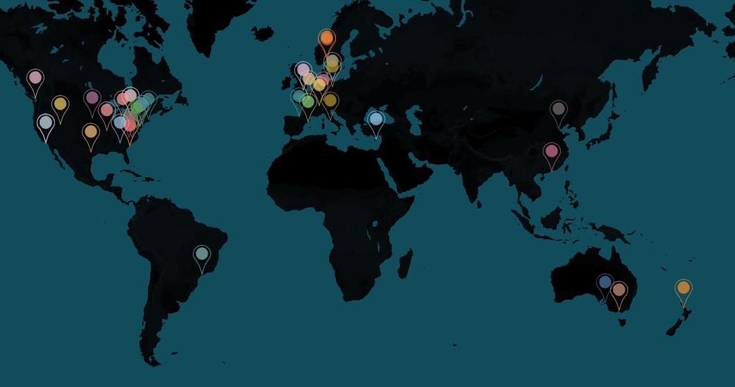
Locations of ENIGMA-Stroke Recovery Members

ENIGMA on social media:


