About ENIGMA-Epilepsy | Secondary Projects | Protocols | Raw Data Analysis: Locipe | Members Page | Join ENIGMA-Epilepsy
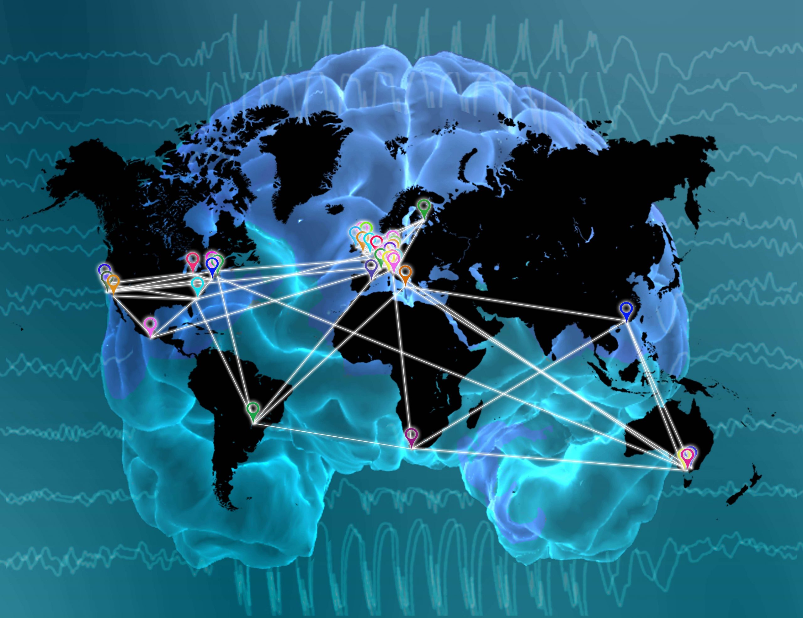
Locations of ENIGMA-Epilepsy Members
 |
Cohorts across:
Chairs: |
[1] The need for standardized MRI meta and mega-analyses in epilepsy
Epilepsy is a neurological disorder characterized by an enduring predisposition to generate epileptic seizures. The disorder may be defined by any of the following: (1) at least two unprovoked seizures occurring more than 24 hours apart; (2) one unprovoked seizure, with a high probability (>60%) of additional, recurrent seizures; (3) diagnosis of an epilepsy syndrome, even if the risk of further seizures is low (for example, benign epilepsy with centro-temporal spikes, BECTS). The International League Against Epilepsy (ILAE) has formally delineated several classifications of seizures and epilepsy sub-types. These classifications have been revised from earlier definitions (see: ILAE reports from 1981 and 1989) in order to improve our organization of recognized forms of epilepsy and facilitate identification of new forms of the disorder. Despite amending these terminologies to include electroclinical characteristics, structural lesions and metabolic abnormalities, our understanding of the neurobiological processes contributing towards, and occurring as a consequence of, epilepsy often remains unclear.
Advances in magnetic resonance imaging (MRI) have helped clinical epileptologists and research scientists study the structure and function of the living human brain in epilepsy in unprecedented detail. For clinicians, neuroimaging now plays a crucial role in neurosurgical planning, localization of the epileptic area and identification of underlying hippocampal sclerosis, tumors or malformations of cortical development in epileptic disorders. For research scientists, MRI has allowed us to scan relatively small (N=10-100) groups of epilepsy patients and healthy controls and then fit statistical models to these scans in order to probe factors that broadly affect neuroanatomy and function in the disorder. Although these studies have identified numerous structural and functional abnormalities possibly associated with the underlying pathology, their modest statistical power typically leads to inconsistent findings from one investigation to the next – overall limiting our ability to reliably generalize any neurobiological insights to the wider population of people with epilepsy.
Some groups have employed systematic reviews, or meta-analyses, of previous neuroimaging studies in order to address these inconsistencies. In these meta-analyses, findings from several independent MRI investigations are reviewed and quantified using ‘random’ and ‘mixed-effects’ statistical models in order to provide more reliable illustrations of underlying biology. Although several meta-analyses have successfully identified more consistent patterns of gray matter atrophy, gray matter hypertrophy and microstructural white matter alteration in epilepsy, they still base their findings on summary statistics from small neuroimaging studies that vary greatly with respect to the processing algorithms and statistical models used on their respective individual datasets. This likely contributes towards confounding inflations of any observed effects, potential ‘blurring’ of more subtle effects, and possible discrepancies from one meta-analysis to the next.
The ENIGMA model of MRI meta-analysis, whereby several independent research groups conduct harmonized neuroimaging analyses on their respective collections of MRI scans, may overcome previous limitations of the meta-analytical paradigm. The ENIGMA model combines important quantitative measures of brain shape, volume and wiring from international research centres, with each centre analysing their own MRI data using identical, standardized protocols. These protocols, which are all freely available online, facilitate efficient analysis of MRI data on large datasets (e.g. up to 31,000 subjects – 79% healthy controls, 21% psychiatric/neurological patients) and have previously been applied in landmark studies of the hippocampus (published in Nature Genetics, 2012), white matter degeneration (published in NeuroImage, 2013) and subcortical brain structures (published in Nature, 2015). This meta-analytic approach may yield sufficient statistical power to robustly and reliably explore neurobiological substrates of epilepsy that no individual site could detect on its own.
[2] ENIGMA-Epilepsy: Overview, aims & objectives of meta-analyses
The ENIGMA-Epilepsy collaboration follows on from other ongoing disease-based ENIGMA working groups such as ENIGMA-schizophrenia, ENIGMA-bipolar, ENIGMA-major depressive disorder, ENIGMA-addiction, ENIGMA-ADD, ENIGMA-HIV, ENIGMA-OCD, ENIGMA-PTSD, and ENIGMA-22q11DS. A broad overview of the ENIGMA-Epilepsy is provided below:
- STAGE 1: Collaborations are formed between epilepsy centers; centers are invited to join ENIGMA-Epilepsy
- STAGE 2: Neuroimaging analysis protocols are circulated to each epilepsy center
- STAGE 3: Analysts at each centre generate a series of standardized statistical models in order to extract summary measures (p-values, z-scores, etc.) quantifying differences in the volume, shape and wiring of various brain structures in people with epilepsy and healthy controls. N.B.: Summary measures are fully anonymized.
- STAGE 4: Summary measures are forwarded to a central meta-analysis site, where they undergo fixed-effects meta-analysis. Meta-analysis may reveal patterns of gray matter damage or white matter loss in particular pre-specified groups of people with epilepsy when compared to neurologically healthy controls.
The overarching aim of the ENIGMA-EPILEPSY collaboration is to improve our understanding of in vivo neuroanatomical disruptions in people with epilepsy compared to the general population. Other important objectives of the collaboration include:
- To create a worldwide network of epilepsy neuroimaging centres.
- To collect summary statistics on brain shape, brain volume and brain wiring from thousands of people with epilepsy and thousands of neurologically healthy control participants in order to vastly improve the statistical power of neuroimaging research.
- To compare and contrast these measures in affected/unaffected groups and, accordingly, illustrate possible differences between the two.
- To identify structural differences between the major forms of epilepsy and major types of seizure.
- To develop collaborations and infrastructure for future analyses
[3] ENIGMA-Epilepsy: Overview, aims & objectives of mega-analyses
The ENIGMA-Epilepsy collaboration follows on from other ongoing disease-based ENIGMA working groups such as ENIGMA-schizophrenia, ENIGMA-bipolar, ENIGMA-major depressive disorder, ENIGMA-addiction, ENIGMA-ADD, ENIGMA-HIV, ENIGMA-OCD, ENIGMA-PTSD, and ENIGMA-22q11DS. A broad overview of the ENIGMA-Epilepsy is provided below:
- STAGE 1: Collaborations are formed between epilepsy centers; centers are invited to join ENIGMA-Epilepsy
- STAGE 2: Data form the different centers are loaded through a password protected web link
- STAGE 3: Stored Data are the pre-processed using our pipeline locipipe
The overarching aim of the ENIGMA-EPILEPSY collaboration is to improve our understanding of in vivo neuroanatomical disruptions in people with epilepsy compared to the general population. Other important objectives of the collaboration include:
- To create a worldwide network of epilepsy neuroimaging centres.
- To collect summary statistics on brain shape, brain volume and brain wiring from thousands of people with epilepsy and thousands of neurologically healthy control participants in order to vastly improve the statistical power of neuroimaging research.
- To compare and contrast these measures in affected/unaffected groups and, accordingly, illustrate possible differences between the two.
- To identify structural differences between the major forms of epilepsy and major types of seizure.
- To develop collaborations and infrastructure for future analyses


